चित्र:Anaplastic astrocytoma.jpg
इस पूर्वावलोकन का आकार: 800 × 552 पिक्सेल। दूसरे रेसोल्यूशन्स: 320 × 221 पिक्सेल | 640 × 442 पिक्सेल | 1,024 × 707 पिक्सेल | 1,200 × 828 पिक्सेल।
मूल चित्र ((1,200 × 828 पिक्सेल, फ़ाइल का आकार: 200 KB, MIME प्रकार: image/jpeg))
|
|
यह फ़ाइल विकिमेडिया कॉमन्स से है। वहाँ पर इसका विवरण पृष्ठ निम्नोक्त है। कॉमन्स मुक्त लाइसेंसों के अंतर्गत उपलब्ध मीडिया फ़ाइलों का संग्रह है। आप भी इसमें मदद कर सकते हैं। |
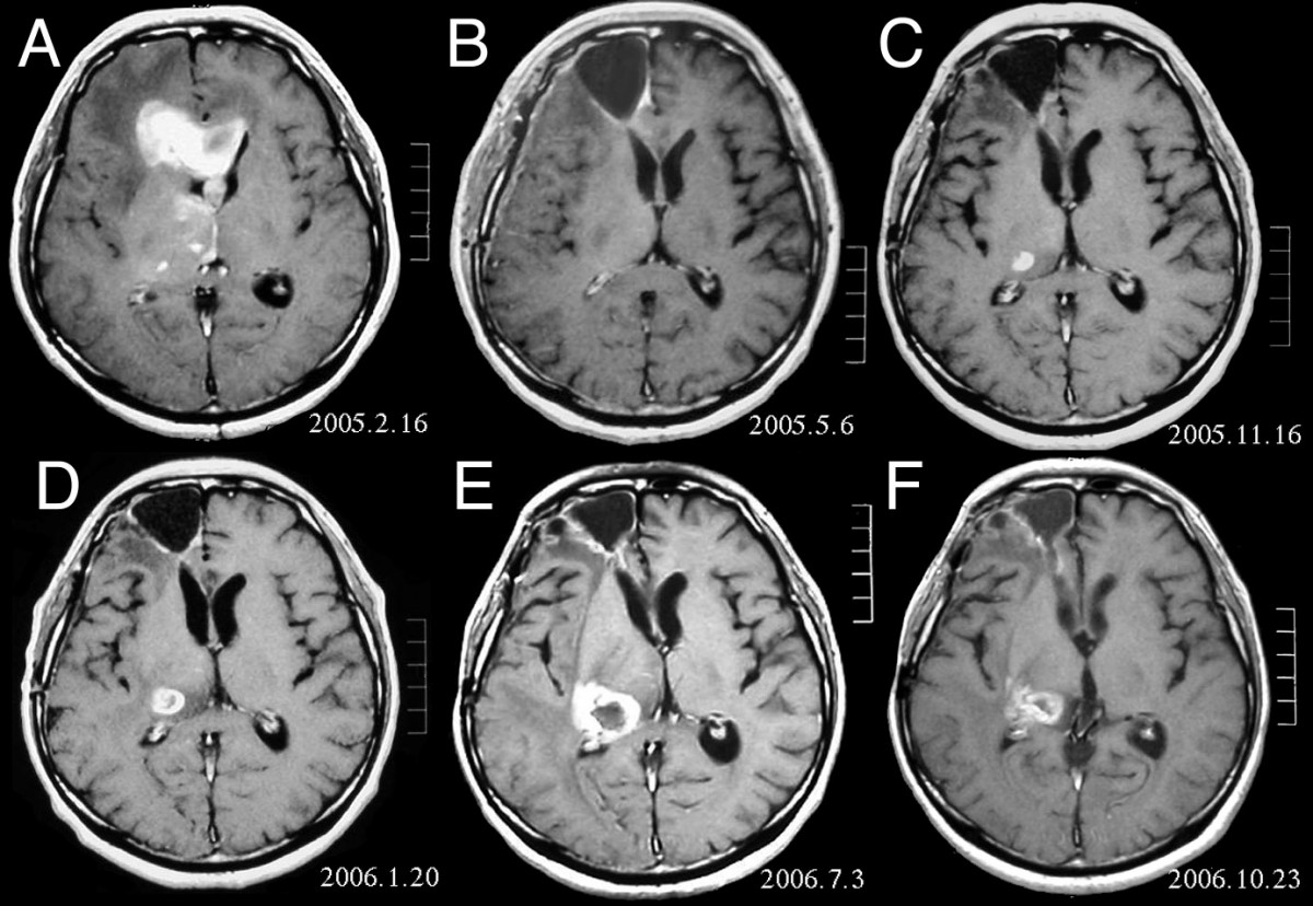
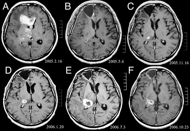
| विवरण | MRI of brain. (A) Initial MRI on February 16, 2005, shows a tumor in the right and left frontal lobe as well as the right thalamus. (B) MRI after surgery, radiation and chemotherapy. The tumor has completely disappeared except for slight enhancement adjacent to the surgical margin. (C) Recurrence of the thalamic tumor despite maintenance chemotherapy on November 16, 2005. (D) Increase in size of the thalamic tumor two months after stereotactic radiotherapy. (E) After 6 cycles of TMZ therapy, the thalamic lesion enlarged, and the patient developed dysarthria and hemiparesis. (F) After 2 courses of treatment with interferon-beta and TMZ, the tumor shows a partial response. |
| दिनांक | |
| स्रोत | Fujimaki T, Ishii H, Matsuno A, Arai H, Nakagomi T.Effectiveness of interferon-beta and temozolomide combination therapy against temozolomide-refractory recurrent anaplastic astrocytoma.World J Surg Oncol. 2007 Aug 4;5:89. PMID 17683572 doi:10.1186/1477-7819-5-89 |
| लेखक | Fujimaki T, Ishii H, Matsuno A, Arai H, Nakagomi T. |
| अनुमति (इस चित्र का पुनः उपयोग करना) |
BioMedCentral License |
इस फ़ाइल को क्रिएटिव कॉमन्स श्रेय 2.0 साधारण लाइसेंस के अंतर्गत लाइसेंस किया गया है।
- आप खुलकर:
- बाँट सकते हैं – रचना की प्रतिलिपि बना सकते हैं, बाँँट सकते हैं और संचारित कर सकते हैं
- रीमिक्स कर सकते हैं – कार्य को अनुकूलित कर सकते हैं
- निम्नलिखित शर्तों के अंतर्गत:
- श्रेय – यह अनिवार्य है कि आप यथोचित श्रेय प्रदान करें, लाइसेंस की कड़ी प्रदान करें, और अगर कोई बदलाव हुए हों तो उन्हें इंगित करें। आप ऐसा किसी भी उचित तरीके से कर सकते हैं, लेकिन किसी भी तरह उससे यह नहीं संकेत नहीं किया जाना चाहिए कि लाइसेंसधारी द्वारा आपको अथवा आपके इस प्रयोग का समर्थन किया जा रहा हो।
चित्र का इतिहास
फ़ाइलका पुराना अवतरण देखने के लिये दिनांक/समय पर क्लिक करें।
| दिनांक/समय | थंबनेल | आकार | सदस्य | प्रतिक्रिया | |
|---|---|---|---|---|---|
| वर्तमान | 16:47, 25 फ़रवरी 2008 | | 1,200 × 828 (200 KB) | Filip em | {{Information |Description=MRI of brain. (A) Initial MRI on February 16, 2005, shows a tumor in the right and left frontal lobe as well as the right thalamus. (B) MRI after surgery, radiation and chemotherapy. The tumor has completely disappeared except f |
चित्र का उपयोग
निम्नलिखित पन्ने इस चित्र से जुडते हैं :
चित्र का वैश्विक उपयोग
इस चित्र का उपयोग इन दूसरे विकियों में किया जाता है:
- ar.wikipedia.org पर उपयोग
- bg.wikipedia.org पर उपयोग
- cs.wikipedia.org पर उपयोग
- da.wikipedia.org पर उपयोग
- de.wikipedia.org पर उपयोग
- el.wikipedia.org पर उपयोग
- en.wikipedia.org पर उपयोग
- es.wikipedia.org पर उपयोग
- et.wikipedia.org पर उपयोग
- hr.wikipedia.org पर उपयोग
- hu.wikipedia.org पर उपयोग
- it.wikipedia.org पर उपयोग
- kk.wikipedia.org पर उपयोग
- lb.wikipedia.org पर उपयोग
- lv.wikipedia.org पर उपयोग
- mk.wikipedia.org पर उपयोग
- mt.wikipedia.org पर उपयोग
- nl.wikipedia.org पर उपयोग
- no.wikipedia.org पर उपयोग
- pl.wikipedia.org पर उपयोग
- ro.wikipedia.org पर उपयोग
- sk.wikipedia.org पर उपयोग
- sl.wikipedia.org पर उपयोग
- sq.wikipedia.org पर उपयोग
- sr.wikipedia.org पर उपयोग
- uk.wikipedia.org पर उपयोग